Embark on a transformative radiology journey with our top-notch courses. Whether you’re a seasoned radiographer or a healthcare professional, our comprehensive programs cater to all levels. Led by industry experts, our personalized guidance, hands-on training, and advanced technologies empower you to excel in modern radiology.
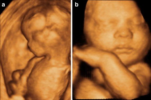
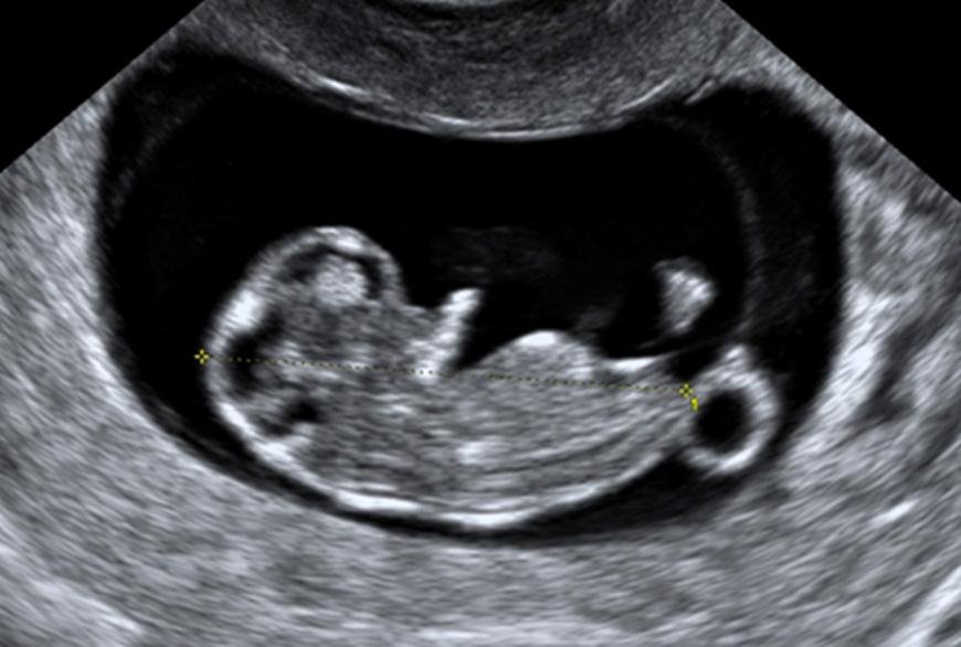
3D-4D obstetric ultrasound provides detailed, lifelike images of the fetus, allowing for enhanced visualization of fetal features and movements in real-time, offering valuable insights for prenatal care and diagnosis.
AVF Doppler evaluation utilizes ultrasound to assess blood flow velocity and direction in the arteriovenous fistula, crucial for monitoring vascular access in hemodialysis patients.
Advanced Extremity Venous Doppler is a non-invasive ultrasound technique used to evaluate blood flow in the veins of the arms and legs, aiding in the diagnosis of deep vein thrombosis (DVT) and other venous disorders. It provides detailed images and real-time blood flow information to assess vascular health and guide treatment decisions.
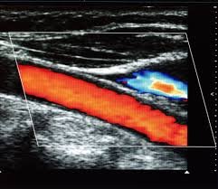
Color Doppler Sonography is a medical imaging technique that visualizes blood flow in real-time, using color-coded maps to indicate direction and velocity. It aids in diagnosing vascular conditions and assessing blood flow dynamics.
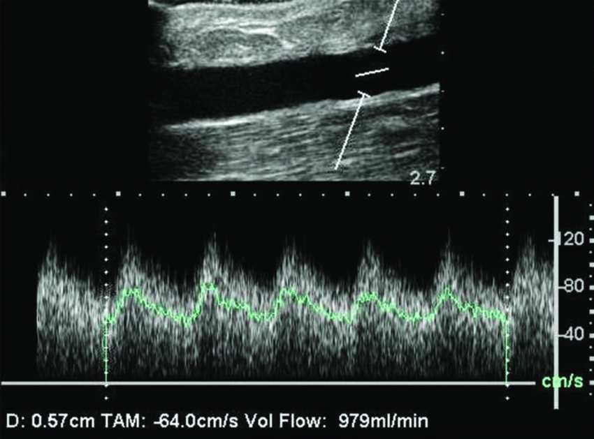
Carotid Doppler is a non-invasive ultrasound technique used to assess blood flow in the carotid arteries, aiding in the detection of blockages or narrowing. Vertebral Doppler similarly evaluates blood flow in the vertebral arteries, crucial for diagnosing conditions like vertebral artery stenosis or dissection.
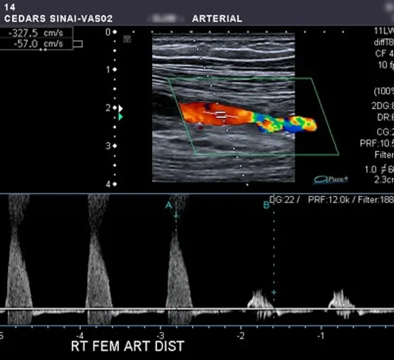
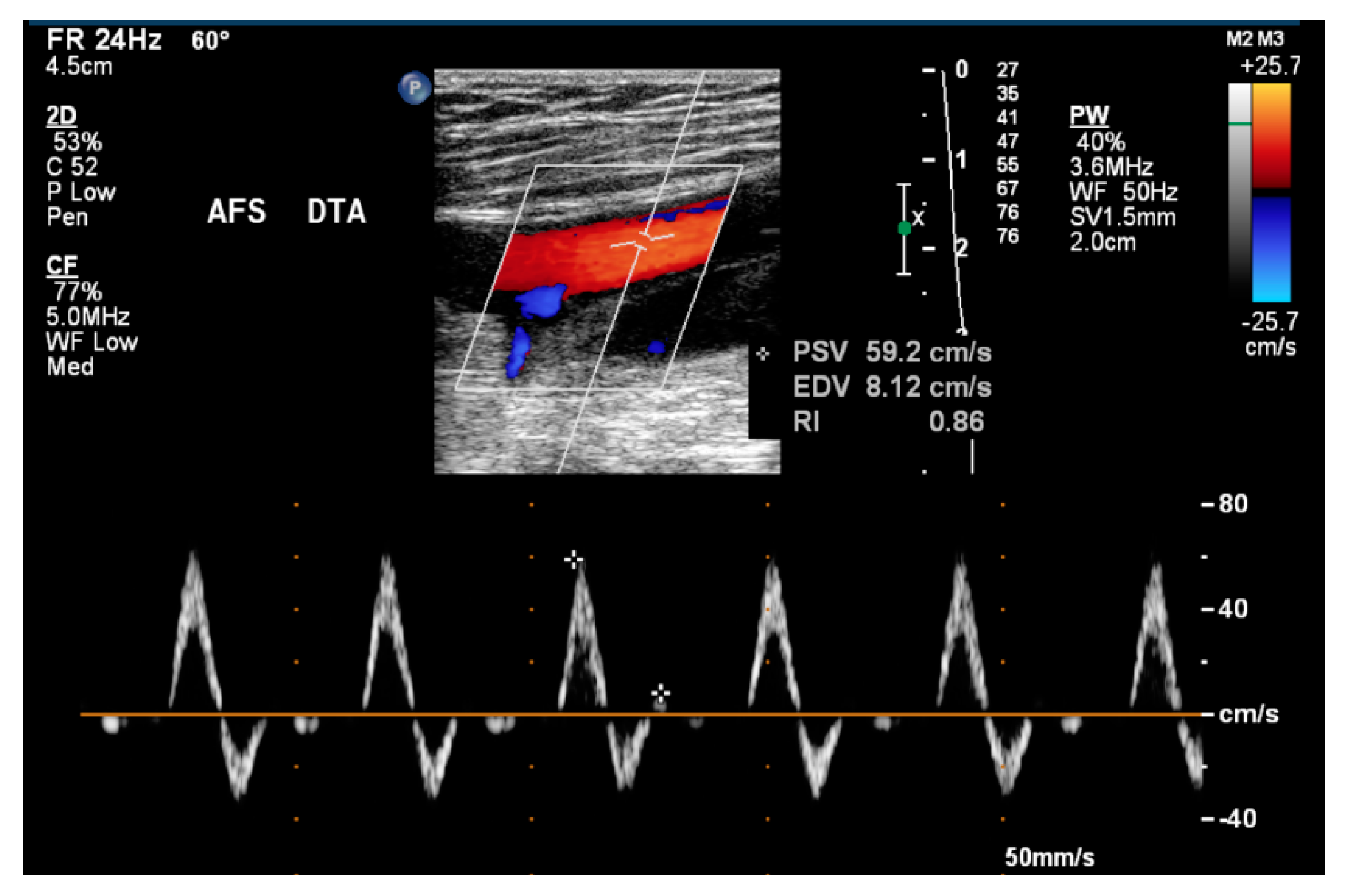
The Advanced Extremity Arterial Doppler is a sophisticated imaging technique used to assess blood flow and detect abnormalities in the arteries of the arms and legs, aiding in the diagnosis of peripheral vascular diseases. It provides detailed insights into circulation, helping clinicians to plan appropriate interventions for patients with vascular issues.
Penile Doppler is a non-invasive ultrasound test used to evaluate blood flow to the penis, aiding in the diagnosis of erectile dysfunction by assessing vascular health and identifying potential causes. It helps determine the effectiveness of blood circulation in achieving and maintaining erections.
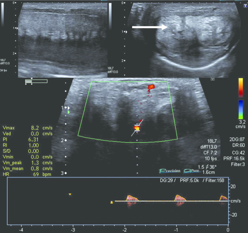
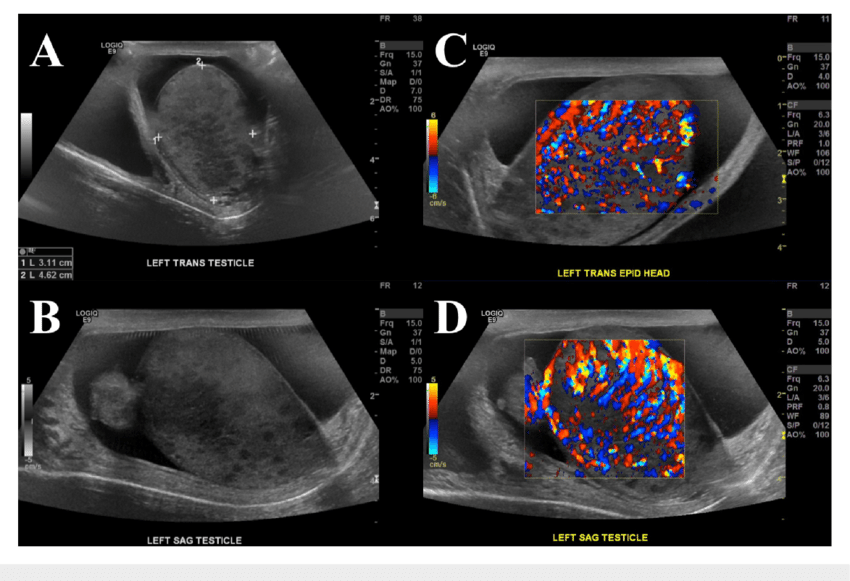
Scrotal ultrasound is a non-invasive imaging technique that uses sound waves to assess the scrotum’s internal structures, aiding in the diagnosis of conditions like testicular torsion or masses. Doppler ultrasound, a variant, assesses blood flow within the scrotum, crucial for evaluating conditions like varicocele or orchitis.
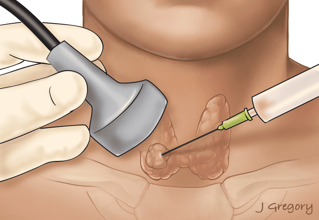
Thyroid biopsy is a diagnostic procedure involving the removal of a small tissue sample from the thyroid gland for examination. Small part biopsy is a minimally invasive technique used to obtain tissue samples from various body parts for diagnostic purposes, aiding in the detection and characterization of abnormalities.
Obstetric ultrasound is a diagnostic imaging technique used during pregnancy to visualize and assess the development of the fetus and monitor maternal health. It helps in determining fetal age, position, and potential complications.
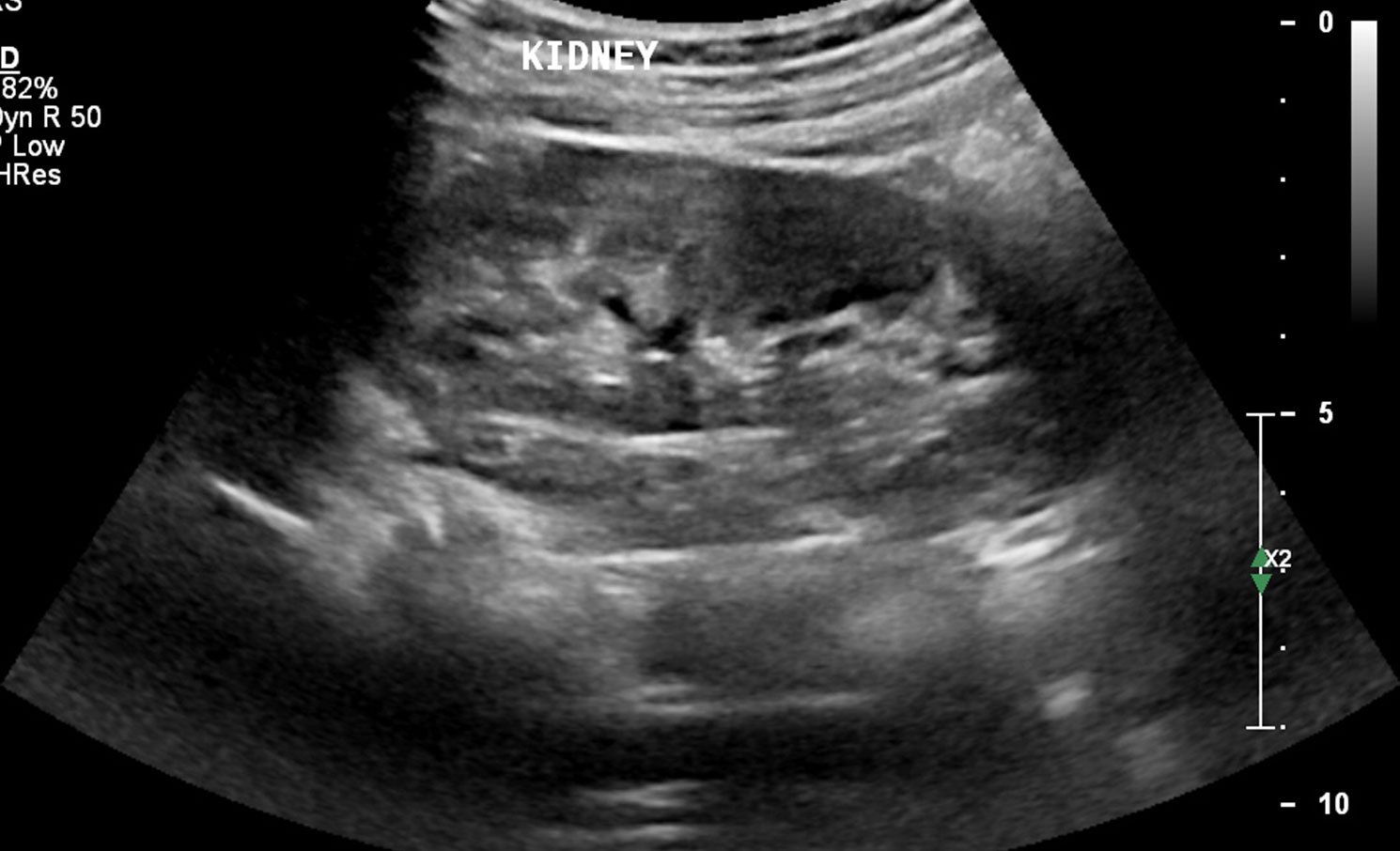
An abdominal ultrasound is a non-invasive imaging technique that uses high-frequency sound waves to produce real-time images of organs and structures within the abdomen. It helps diagnose and monitor conditions related to the liver, kidneys, pancreas, gallbladder, and spleen.
A male pelvic ultrasound is a non-invasive diagnostic imaging procedure used to visualize the organs and structures within the male pelvis, including the bladder, prostate, and seminal vesicles. It helps in assessing conditions such as prostate enlargement, tumors, and urinary disorders.
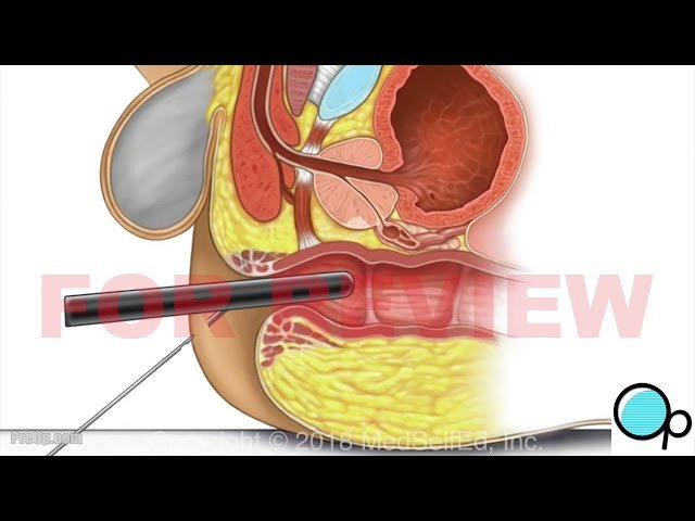
A transrectal prostate ultrasound and biopsy is a diagnostic procedure where an ultrasound probe is inserted into the rectum to create images of the prostate, and small tissue samples are collected with a needle for analysis to detect prostate abnormalities, including cancer.
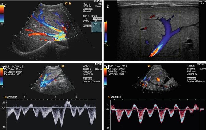
An Abdominal Doppler is a non-invasive ultrasound technique used to visualize the blood flow in the abdominal organs and vessels, helping diagnose conditions related to circulation abnormalities such as blockages and narrowing of arteries.
Understanding 3D Transvaginal Ultrasound involves using advanced imaging technology to create three-dimensional images of the female reproductive system, providing detailed views that assist in diagnosing and managing gynecological conditions.
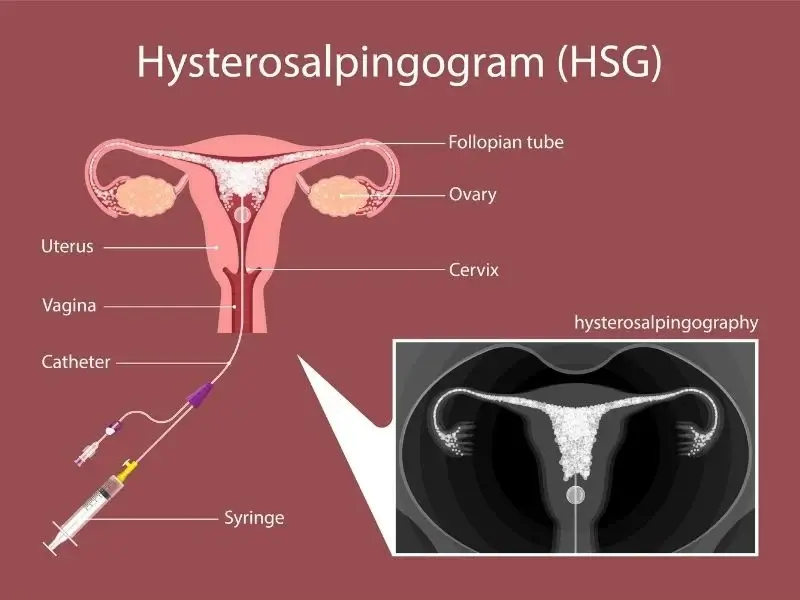
Hysterosalpingography is a radiographic diagnostic procedure that uses a contrast medium to visualize the uterine cavity and fallopian tubes, helping assess their shape and function, and identifying potential blockages or abnormalities that could affect fertility.
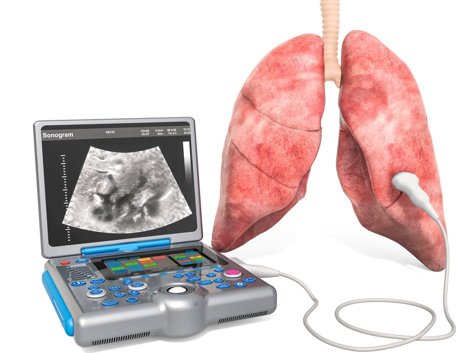
“Understanding Lung Ultrasound” is a comprehensive guide that explores the techniques and applications of lung ultrasound, offering clear, step-by-step instructions to aid healthcare professionals in diagnosing and monitoring pulmonary conditions effectively. This resource emphasizes practical skills and image interpretation, enhancing clinical decision-making in real-time patient care.
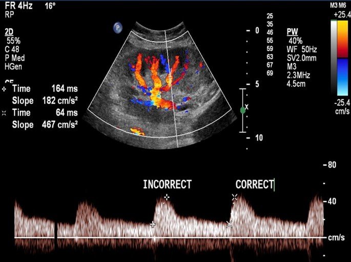
Renal Transplant Doppler is a non-invasive imaging technique used to assess blood flow in transplanted kidneys, aiding in the evaluation of graft function and detecting complications such as rejection or vascular compromise. It utilizes Doppler ultrasound to measure velocity and direction of blood flow within the renal vasculature.